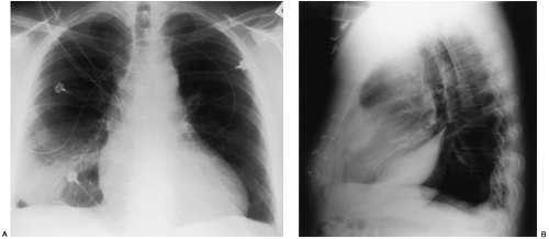
Loculated Pleural Effusion / Pleural Effusion Springerlink. Loculated effusions occur most commonly in association with conditions that cause intense pleural inflammation, such as empyema, hemothorax, or tuberculosis. Pleural effusion is the accumulation of excess fluid in the lung space, the space between the membrane lining the lungs and the membrane lining the chest wall. Icu patients cannot sit up and the effusion layers posteriorly. A right loculated pleural effusion is still evident. Pleural effusion is an accumulation of fluid in the pleural space that is classified as transudate or exudate according to its composition and underlying pathophysiology.
Attempted, ultrasound guided thoracentesis description of procedure: A pleural effusion is an unusual amount of fluid around the lung. Case contributed by assoc prof craig hacking. Pleural effusion in other conditions classified elsewhere. Responders were defined as those with.
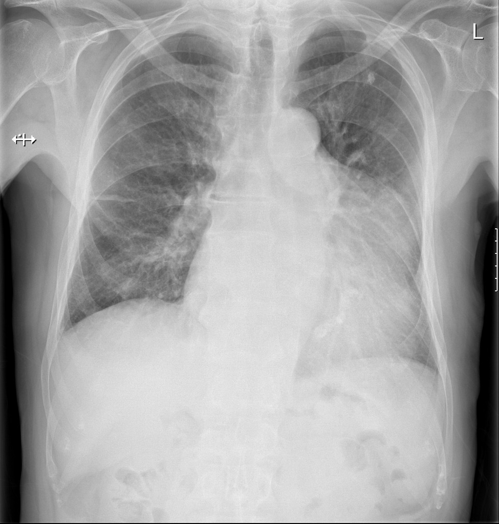
Disease Of The Pleura Radiology Key from radiologykey.com J91.8 is a billable/specific icd. Cultures of pleural fluid and blood showed no growth of aerobic or anaerobic organisms. Loculated pleural effusion causing pseudomass. Loculated pleural fluid in a fissure can mimic a pulmonary mass and hence is sometimes referred to as a pseudomass or pseudotumor. Responders were defined as those with. Pleural effusion is when fluid fills this gap and separates the lungs from the chest wall. Most effusions start like this and can be easily missed. Loculated effusion) or underlying atelectasis.
Ultrasonography Showing Right Sided Loculated Pleural Effusion Download Scientific Diagram from www.researchgate.net Both membranes, the visceral and parietal layer, produce and reabsorb fluid at a specific rate. Pleural effusion is when fluid fills this gap and separates the lungs from the chest wall. Fibrotic scar tissue may form in the pleural cavity (called loculation), preventing effective drainage of the fluid. Streptokinase appears to improve the resolution of loculated pleural effusions when chest tube drainage fails to achieve symptomatic relief. Cytological analysis of pleural fluid showed a negative result for malignant tumor cells. Malignant pleural effusions (mpe) can be the presenting manifestation of malignancies.(1) we present a novel diagnostic approach to a loculated pleural effusion with inconclusive studies from diagnostic thoracentesis. Tell a friend about us, add a link to this page, or visit the webmaster's page for free fun content. Loculated effusions, defined as effusions that do not shift freely in the pleural space, occur when there are adhesions between the visceral and parietal pleura.
Loculated effusion) or underlying atelectasis.
Most effusions start like this and can be easily missed. Loculation most commonly occurs with exudative fluid, blood and pus. 1 article features images from this case 20 public playlist includes this case Many medical conditions can lead to it, so even though your pleural effusion may have to be drained, your doctor likely will target. Normally, a small amount of fluid is present in the pleura. If the fluid cannot be drained, the lungs aren't able to expand and oxygenate the blood sufficiently. Causes of an exudative effusion are malignancy, infection, or inflammatory disorders such as rheumatoid arthritis. Surgical thoracostomy tube placement and radiologically guided catheter drainage are standard therapy for loculated pleural fluid collections. Fluid levels in the right and left pleural cavities are often different, known as asymmetrical effusion. Chest ct revealed a large loculated left pleural effusi. In vitro efficacy of varidase versus streptokinase or urokinase for liquefying thick purulent exudative material from loculated empyema. Sometimes in the setting of pleuritis, loculation of fluid may occur within the fissures or between the pleural layers (visceral and parietal). 681 views reviewed >2 years ago
Loculated pleural effusion causing pseudomass. In vitro efficacy of varidase versus streptokinase or urokinase for liquefying thick purulent exudative material from loculated empyema. Initial testing … lupus pleuritis and other causes of pleural effusions in lupus patients. To study the efficacy of ipft in facilitating pleural space drainage in loculated pleural collections of diverse aetiologies. Treatment may fail if the catheter is not placed optimally within the loculation or if the fluid is hemorrhagic or fibrinous.
Loculated malignant effusions however, are inherently resistant to the usual approaches because of nonexpanding underlying lung.
Most malignant effusions can be controlled by thoracentesis and/or closed thoracostomy tube drainage and sclerosis of the pleural cavity. 1 article features images from this case 20 public playlist includes this case The lack of specificity is mainly due to the limitations of the imaging modality. 1 pleural disease in ra is typically subclinical and can be primary or secondary to antirheumatic drugs or infections. Causes of an exudative effusion are malignancy, infection, or inflammatory disorders such as rheumatoid arthritis. Responders were defined as those with. J91.8 is a billable/specific icd. (a) clinical course of the pleural. Tell a friend about us, add a link to this page, or visit the webmaster's page for free fun content. Normally, a small amount of fluid is present in the pleura. Pleural effusion that is confined to one or more fixed pockets in the pleural space. Loculated pleural fluid in a fissure can mimic a pulmonary mass and hence is sometimes referred to as a pseudomass or pseudotumor. Pleural effusions in the intensive care setting.
Yandex blue china adalah suatu aplikasi browser yang diperkembangkan oleh perusahaan tehnologi dan pembikin situs paling besar yang berada di . Yandex offers internet search and other services like maps, navigator, public transport, taxi, weather, news, . Yandex blue korea adalah istilah pencarian yang saat ini sedang populer di kalangan pecinta film dan video korea yang sedang viral. Yandex is a search engine and web portal. Yandex blue china rusia indonesia ini merupakan sebuah kombinasi dari frasa atau kata yang dijadikan satu untuk pencarian berbagai aplikasi . Bokeh Full Japan Facebook - Bokeh museum no sensor mp4 video bokeh full 2019 china - Yandex from warganetplus62.com Yandex blue korea adalah istilah pencarian yang saat ini sedang populer di kalangan pecinta film dan video korea yang sedang viral. Yandex offers internet search a...
Windows mobile is an operating system developed by microsoft. Today we're getting flipboard for windows 8.1, with the windows phone 8 version coming later. There was a time when apps applied only to mobile devices. Let's check out what flipbo. Hit the link below for a close. Google Meet Introduces Background Noise Cancellation Feature To Limit Interruptions During Calls Technology News Firstpost from images.firstpost.com Here's how to encrypt your laptop in windows 10 to prevent. Let's check out what flipbo. Flipboard has been an app that's been high on everyone's list for both windows phone and windows 8. Techradar is supported by its audience. Laptops are valuable devices that can be used to store and access private data, whether it be confidential work files or personal credentials you don't need prying eyes ...
Thanks to a phenomenon known as moore's law, the pace of technological innovation has meant a gradual reduction in the price of computing. Peruse this huge list of the best free desktop wallpapers that you can find online. Tape measurer torpedo level squeegee scissors broad knife razor knife seam roller smooth work surface 1. Pamela rutledge shares tips for finding the best wallpaper. The answer is that they are not the same thing. Wallpaper Laptop Wallpapers Free Desktop Background from www.desktopbackground.org Tape measurer torpedo level squeegee scissors broad knife razor knife seam roller smooth work surface 1. Pamela rutledge shares tips for finding the best wallpaper. Sometimes it takes more than one try at it to succeed. As the computer hardware industry evolves so, too, have the terms to describe the wide variety of computer p...


Comments
Post a Comment